

The immune system is full of T cells that could hunt down and destroy tumors, if they were primed. Now, a UC San Francisco team has invented a way to do this by combining immune cells that are grown in the lab with samples from a patient’s tumor.
Like giving a scent to a hound dog, it provides direction to the immune system on how to track down the cancer.
To catch the scent, the T cells need to be trained using rare dendritic immune cells that are hard to obtain from cancer patients. The UCSF method gets around this by making dendritic cells from induced pluripotent stem cells (iPSCs), which can be grown in large quantities in the laboratory.
Since typical iPSCs would be rejected by the immune system, the scientists first strip them of identifying information, then prompt them to mature into dendritic cells. Finally, they “dress” the dendritic cells with tiny membrane bubbles made from tumor cells. This gives the dendritic cells many different views of the cancer to present to the T cells, creating a highly complex scent for the hounds to track.
In laboratory experiments using tumor samples and T cells from patients with leukemia and ovarian cancer, the dressed dendritic cells were able to train the T cells to kill tumors from the same patient. The treatment also slowed tumor growth in a mouse model. The findings appear in Cell Stem Cell on Aug. 25.
“For cell-based therapies to work, they need to avoid immune rejection by the body, and they need to target the cancer and not healthy tissue,” said Robert Blelloch, MD, PhD, a professor in the Department of Urology at UCSF and senior author of the paper. “Our combination of iPSC-derived dendritic cells with patients’ own tumor signature checks both boxes and we’re very hopeful it could work in the clinic.”
A new immunotherapy for cancer
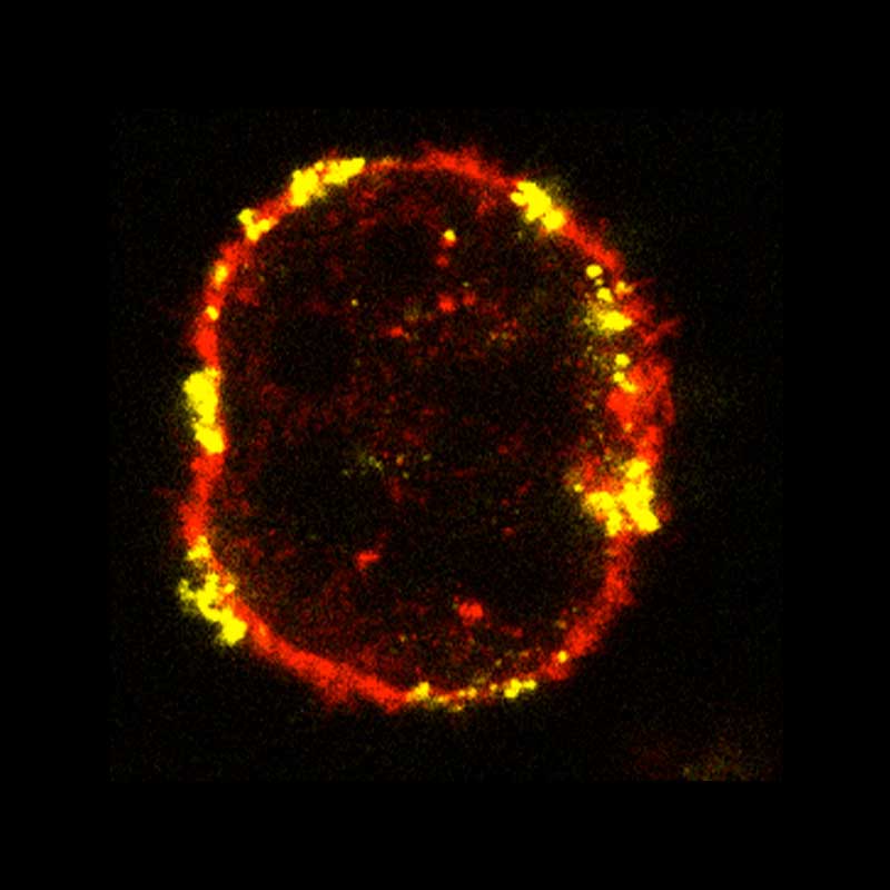
Lab-grown dendritic cells (red), which train the immune system to destroy pathogens or infected cells, were exposed to bits of tumor cells (yellow) taken from a tumor biopsy. The dendritic cells absorbed the bits of the tumors into their own surface. The UCSF team showed these engineered immune teachers could educate killer T-cells to find tumor cells in a mouse model.
Credit: Xu et al., Cell Stem Cell.
Home-schooling the immune system
Instead of trying to identify the right cancer targets one by one, the researchers broke apart the membranes of tumor cells and let them form into tiny bubbles. These bubbles merged with the dendritic cells, coating them with tiny pieces of the patient’s tumor.
This both made the dendritic cells look more like the patient’s own cells and carried a wide range of cancer targets. All that information could make it harder for a tumor to escape just by shedding a single target, a problem that can limit other immunotherapies.
Once the scientists showed that the tumor membrane had integrated into the dendritic cells, they tested whether the cells could prompt T cells to find and fight cancer.
The dendritic cells did more than carry pieces of the tumor. They also produced the specific instructions that the T cells needed to recognize the tumor pieces as a threat — and the scientists were able to amplify those “attack” instructions, leading to an even stronger T cell response.
The approach worked in petri dishes using tumor samples and T cells from the same patients, and it slowed the growth of human cancer cells in mice.
For Blelloch, one potential use is easy to imagine. Surgery can remove prostate tumors, for example, but some patients still face a high risk that the cancer will return.
“After surgery, we have all the tumor tissue needed to make a bespoke therapy for that patient’s cancer,” Blelloch said. “We could dress artificial dendritic cells with pieces of the biopsy, place them back in the patient to educate T cells, and then the T cells could go kill any remaining cancer cells in the body, removing the risk of the tumor returning.”
Authors: Other UCSF authors are Huaigeng Xu, MD, PhD; Chisato Kamei, MD; Deniz Goekbuget, PhD; Jacob Du; Mercedes Barrero, PhD; Li Wang, PhD; Jason Mark Soriano; Serena S. Kwek, PhD; Lawrence Fong, MD; and Karin M.L. Gaensler, MD. For all authors, see the paper.
Funding: This work with supported by the National Institutes of Health (U01CA244452), the California Institute of Regenerative Medicine (DISC0-13806, EDUC4- 12812), a NIH Cancer Moonshot Award, a Sandler Program for Breakthrough Biomedical Research New Frontiers Award, and Benioff Initiative for Prostate Cancer Research.