

A new AI-based model can predict where the most common malignant brain cancer is most likely to return after initial surgery, potentially giving doctors a chance to treat it before it becomes visible on MRI.
Glioblastoma is the most lethal brain tumor in adults, with a median survival of about 17 months after diagnosis. Even after surgeons remove all visible tumor and patients receive follow-up treatment, the cancer almost always returns.
Now, researchers led by UC San Francisco and University of Michigan have developed an AI approach that can predict, with a high degree of accuracy, where first recurrence is likely to appear. In glioblastoma, most patients experience recurrence at or close to the tumor cavity.
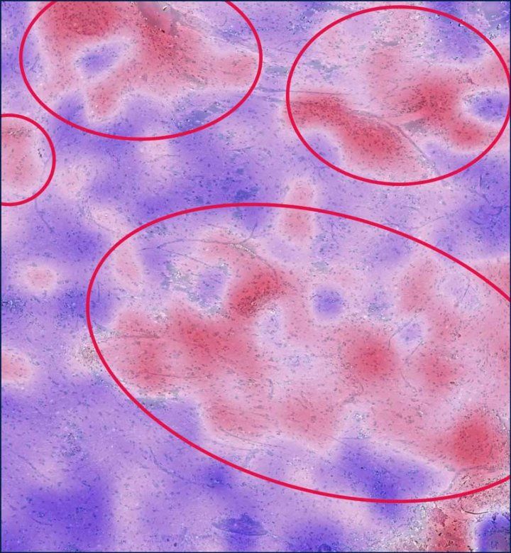
The findings, published Sept. 25 in Science Advances and supported by the National Institutes of Health (NIH), could eventually enable doctors to use targeted treatments in these areas before a new tumor becomes visible on MRI.
“The system has incredible potential to provide neurosurgeons with valuable real-time guidance during tumor removal,” said first author Sanjeev Herr, MD, a postdoctoral research fellow at UCSF. “It can also generate insights that guide subsequent treatment decisions.”
These treatments might include removing additional tissue during the initial surgery when it is safe to do so, said senior author Shawn Hervey-Jumper, MD, a neurosurgeon at UCSF Health, and Mitchel S. Berger, MD, endowed professor at the Weill Institute for Neurosciences.
“Patients with disease in parts of the brain that cannot be removed may undergo other treatments targeted at the predicted sites of progression, like higher-dose focal radiation or drugs infused directly into the tumor, via a catheter placed through the skull,” Hervey-Jumper said.
AI analyzes tissue samples for migrating cancer cells
To develop the model, the researchers analyzed tissue samples collected during glioblastoma surgery from UCSF Health patients, for whom the median time to glioblastoma recurrence was 5.5 months. Their model used about 300 samples from 60 patients and was tested separately on about 100 samples from another 20 patients.
The researchers produced microscopic images from these tissue samples, which they analyzed with Fast Glioma, an AI system developed by UCSF and University of Michigan. Tissues were scored based on tumor infiltration, which is the presence of cancer cells that had migrated to healthy surrounding areas.
The AI score alone performed about as well as conventional pathology at predicting which areas would later develop recurrent tumor. But when researchers combined the AI score with clinical, imaging, and molecular data that had been tested on six machine-learning models, they found that one model was significantly more likely to distinguish between sites that would and would not develop recurrences.
The researchers also tested how precisely the model could pinpoint where recurrence would develop. The model performed well at predicting whether cancer would return within 5 or 10 millimeters of the tissue that had been sampled.
The team chose the first recurrence because patients often receive experimental treatments later in their disease that can influence tumor growth, making subsequent recurrences more difficult to predict reliably.
“The overall goal was to delay that first recurrence,” said co-senior author Todd Hollon, MD, of the Machine Learning in Neurosurgery Laboratory at the University of Michigan, Ann Arbor. “Ultimately, we hope that extending that window could translate into longer survival.”
Authors: Please see the study.
Funding: National Institutes of Health (NINDS R01 NS137950, K12NS080223, T32GM007863, NCI P01 CA118816), and additional foundation, philanthropic and institutional sources. There are no disclosures to report.