
When you reach into a bucket of ice, step outside on a snowy day, or feel the tingle of menthol toothpaste, a protein in your nerve cells called TRPM8 springs into action, opening like a tiny gate to send a “cold” signal to your brain.
For years, scientists have been trying to figure out how that gate changes as it opens, in hopes of using it as an entry point for future pain therapies.
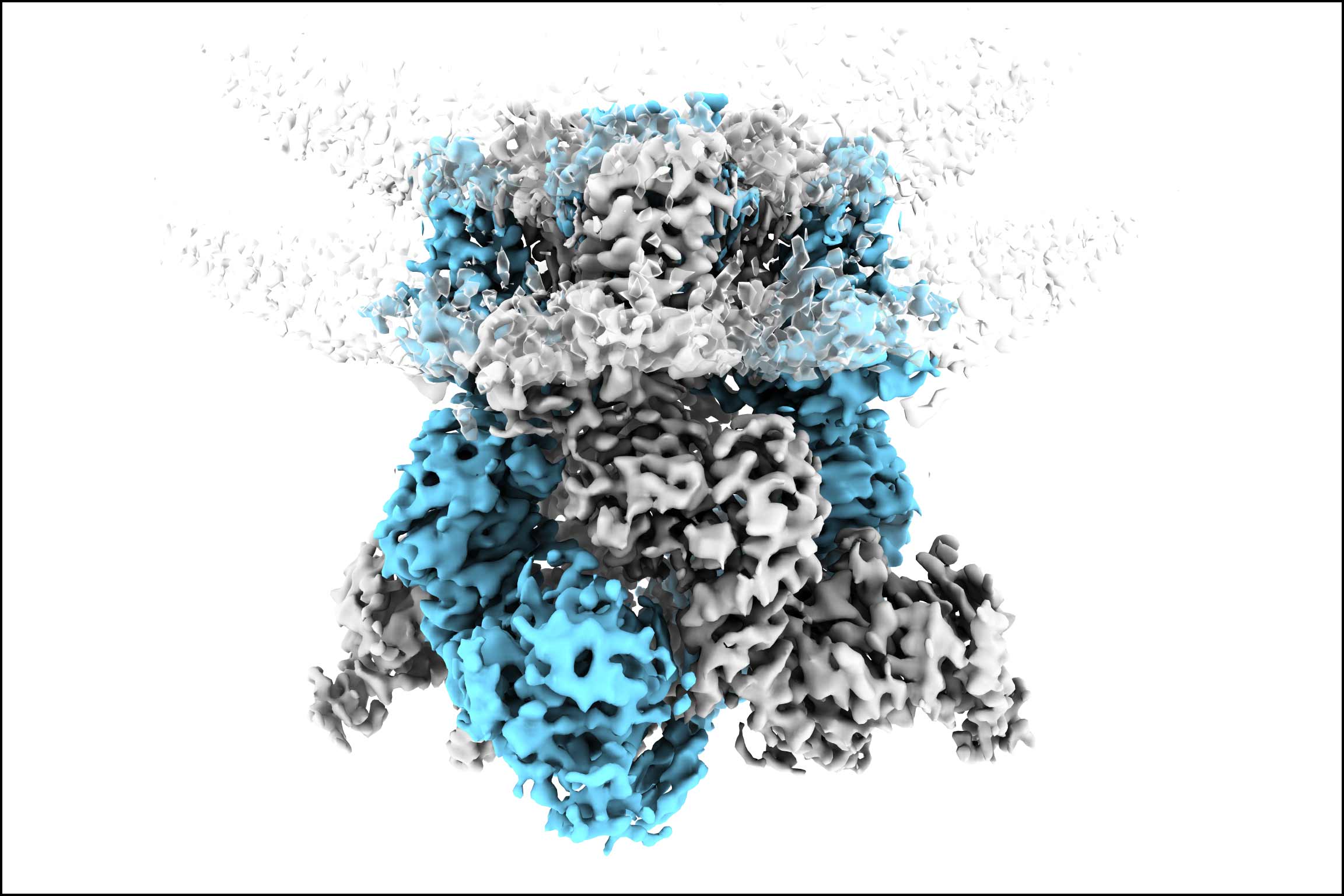
Now, UC San Francisco researchers have discovered exactly how TRPM8 changes its shape as temperatures cool, enabling them to create a three-dimensional picture. The work, published in Nature on March 25, answers an important question in pain research. It also explains why birds — which also have TRPM8 in their nerve cells — are far less sensitive to cold than mammals.
“Everyone always wants to know how temperature sensing works, but it turns out to be a very technically challenging question to answer,” said co-senior author David Julius, PhD, chair of Physiology and the Morris Herzstein Chair in Molecular Biology and Medicine, both at UCSF. “So, to finally have insight into this is really very exciting.”
Julius received the 2021 Nobel Prize in Physiology or Medicine for discovering TRPV1, which enables nerves to sense capsaicin, the spicy heat of chili peppers.
A key to the cold discovery was being able to see proteins in motion.
“For decades, structural biology has focused on capturing proteins in stable, frozen states. This work shows that to truly understand how a protein functions, you also have to understand how it moves,” added Yifan Cheng, PhD, UCSF professor of biochemistry and biophysics and an investigator at the Howard Hughes Medical Institute (HHMI) who co-led the work.
A stubborn protein
Scientists knew that TRPM8 begins to activate when temperatures dip below about 79 degrees Fahrenheit. Yet despite years of effort, researchers had been unable to capture its exact molecular structure while responding to cold.
Most imaging methods also rely on proteins being locked in a single, stable structure to visualize them — limiting scientists’ ability to see fluid, intermediate structures as a protein changes shape. Yet TRPM8, which is normally embedded in the membrane of nerve cells, tended to fall apart when isolated.
Julius’ and Cheng’s teams solved this by imaging TRPM8 while it was still embedded in membranes that were taken directly from cells.
“We realized that the protein is particularly sensitive to how you handle it. Keeping it in the native membrane was what finally let us see what was actually happening,” said Kevin Choi, a graduate student at UCSF and co-first author of the study.
Mapping the effect of cold
To capture TRPM8 as it opened, the team used two complementary techniques: cryo-electron microscopy (cryo-EM), which takes still pictures, and hydrogen-deuterium exchange mass spectrometry (HDX-MS), which is more dynamic.
For cryo-EM, they prepared samples of the protein in cold, with menthol, or at room temperature. Then, they flash froze the samples. This locked the channel into its shape at that moment. Cryo-EM then generated 3D snapshots of the protein's atomic arrangement.
They used HDX-MS to track the protein in real time as the surrounding temperature changed. The method highlighted which regions of the molecule flex and move as the temperature changed. Together, the methods let the researchers model exactly how TRPM8 opened below 79 degrees.
“Just as looking at a photo of a horse can’t tell you how fast it runs, the electron microscopy alone can’t tell us how the molecule moves and what drives those movements,” said co-first author Xiaoxuan Lin, PhD, an HHMI staff scientist working in Cheng’s lab at UCSF. “But combining these two techniques gave us a window into what was happening.”
The analysis revealed that cold stabilizes a specific region of the TRPM8 channel, which triggers a key helix to move. This enables a separate lipid molecule to slide into that spot, locking the channel open and sustaining the cold signal.
When the researchers compared human TRPM8 with the bird version of the protein, which responds to menthol but is far less cold-sensitive, they were able to detect which features are specifically responsible for detecting cold.
A lesson for structural biology
The new work paves the way for determining the structure of other dynamic proteins that have typically been hard to image.
“The lessons we learned in studying this channel are actually very broadly useful,” Cheng said. “Dynamic behavior is critical for the function of many proteins, and you can’t understand dynamic behavior from one snapshot of a protein’s structure.”
Julius and Cheng are now applying the same strategy to get a better understanding of TRPV1, the heat-sensing channel that Julius discovered in 1997. They also plan to examine how compounds that block TRPM8 — several of which are in clinical trials for pain — affect the structure of the protein. Ultimately, that could lead to more targeted treatments for conditions like cold allodynia, in which even mild cold triggers severe pain.
Funding: This work was supported by grants from the NIH (R35NS105038 and R35GM140847). Instruments at the UCSF Cryo-EM facility are partially supported by grants from the NIH (S10OD020054, S10OD021741 and S10OD026881) and Howard Hughes Medical Institute.
Disclosures: Cheng serves on the scientific advisory boards for ShuiMu BioSciences and Pamplona Therapeutic Co.