Cellular response to stress signals predicts future tumor formation in women diagnosed with common

A specific biological response to cellular stress may predict the likelihood of future tumor formation of the most common, non-invasive form of pre-malignant breast cancer—ductal carcinoma in situ, or DCIS.
This information could potentially be used in a clinical setting to determine which women should receive more or less aggressive therapy when initially diagnosed with DCIS, according to a study led by researchers from the University of California, San Francisco.
The research results are also significant because traditional tests available today are not strong enough to predict whether or not a woman will develop a future cancer after diagnosis with DCIS. By identifying this particular biological response in patients, physicians may now be able to predict subsequent tumor formation years before it actually occurs.
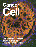
The study is the cover story of the November 13, 2007 issue of “Cancer Cell.” It is a translational study that brings together an integrated team of basic and clinical scientists in an attempt to find molecular markers to aid women and their physicians in determining the best treatment option following a diagnosis of DCIS.
“We were very excited by the results,” said senior author Thea Tlsty, PhD, professor of pathology and co-leader of the Cell Cycling and Signaling Program of the UCSF Comprehensive Cancer Center. “Until now, little has been known about the molecular pathways that may cause a differential risk in women diagnosed with this type of breast pre- malignancy.”
DCIS is a type of breast cancer where cancer cells form inside the milk ducts of the breast but have not spread to surrounding breast tissue. DCIS accounts for nearly 25 percent of all breast cancer diagnoses, according to the American Cancer Society.
In the absence of this new information, the researchers said, women diagnosed with DCIS today are often offered a gamut of treatment options that extend from a full mastectomy to watchful waiting. A majority of women usually opt for a lumpectomy with or without additional treatments such as radiation, hormonal therapy or chemotherapy. However, since the majority of DCIS lesions are not associated with formation of subsequent invasive tumors, it is likely that many women diagnosed with DCIS are being over treated.
“Conversely, since some women still get subsequent invasive carcinomas following these therapies, we may be under-treating a group of women diagnosed with DCIS,” said Tlsty. Approximately 12-15 percent of women diagnosed with DCIS develop a subsequent invasive tumor within 10 years after undergoing surgical lumpectomy.
As noted in the study, it is well recognized that normal cellular responses to stress are important barriers to cancer formation and therefore also provide researchers with molecular candidates to help identify lesions that will not progress to a malignancy. To determine if stress-associated biomarkers in this response could provide insight into the likelihood of subsequent tumor formation in DCIS, the researchers examined several biomarkers in biopsies of DCIS tissue both in isolation and in conjunction to determine their effects.
They found that the presence of high amounts of two biomarkers, p16 and COX-2, played a critical role in the biological response to cellular stress. In particular, they found that tumors with high p16 and/or high COX-2 in the absence of cell proliferation reflected correct stress activation and a protective response to cellular stress.
“This phenotype begins to identify women less likely to have a subsequent tumor event after a diagnosis of DCIS,” said Tlsty. “It supports the hypothesis that the senescent program is a barrier to tumor growth.”
In a second group, DCIS tissue with high p16 and/or high COX-2 in the presence of ongoing cell proliferation corresponded to a short-circuited response to cellular stress. The lesions in this group had lost functional p16/retinoblastoma (Rb) signaling, allowing cells to proliferate unchecked and thus likely to lead to subsequent tumor formation. This finding positively identified the Rb pathway as a key regulator in these responses.
Study findings showed that the expression of biomarkers indicative of the short-circuited response was also a defining characteristic of basal-like invasive tumors, a more aggressive form of breast cancer, which was a striking discovery, according to Tlsty.
“We found that the expression of biomarkers of a stress-associated response is conditional and can reflect two distinct processes distinguished by the absence or presence of a proliferation of cells,” added Tlsty. “We anticipate that these phenotypes have the potential to accurately predict the occurrence of future tumor events in women diagnosed with DCIS.”
The biological phenotypes identified in this study are now being validated in a large, independent study of women diagnosed with DCIS. Initial results are expected within the next year.
This study was supported by the National Cancer Institutes Breast Cancer Specialized Program of Research Excellence (SPORE) grant and the Cancer League.
Study co-leaders were Mona Gauthier, PhD, and Hal Berman, MD, both of the UCSF Department of Pathology, and Karla Kerlikowske, MD, San Francisco VA Medical Center. Additional investigators were Karen Chew, UCSF Comprehensive Cancer Center; Dan Moore, PhD, California Pacific Medical Center; Caroline Miller, Krystyna Kozakiewicz, PhD, Joseph Rabban, MD, and Yunn Yi Chen, MD, PhD all of the UCSF Department of Pathology.